Protein Phycocyanin Structure
This page contains information about accessing structural images of the protein phycocyanin which is the subject of Experiment: Protein Folding and Extraction (CHM 1415, General Chemistry II Lab).
The phycocyanin structure file is available from the Research for Collaborative Structural Bioinformatics, RCSB, which is a worldwide repository for processing and distribution of 3-D biological macromolecular structural data. The RCSB Protein Data Bank is jointly operated by NSF, DOE, and NIH.
You can view the structure directly on the RCSB web site or you can download it to your computer and then view it:
Go to the RCSB homepage. In the search box type 1GH0 which is the id code for phycocyanin. A summary page of the structure that includes a preview image of 'biological assembly 1' as well as additional structural information will display. You can scroll through other preview images which focus of different structural aspects of the protein in this frame using the radio buttons (top) or arrow icons (bottom) of the image.
Select the 3D View tab at the top of the page to view the structure directly in your browser. The default viewer is JSmol which is a Java-based viewer and which some browsers may block from opening without user permission. You can change the viewer from JSmol to NGL or PV using the pull-down menu below the structure image. NGL and PV viewers are WebGL-based viewers and the former is more feature rich.
You can also download the phycocyanin structure file to your computer and view it using a standalone viewer. Links to several free standalone viewers are below the preview image on the summary page. A very robust standalone viewer is Chimera which can be downloaded from the UCSF Chimera webpage.
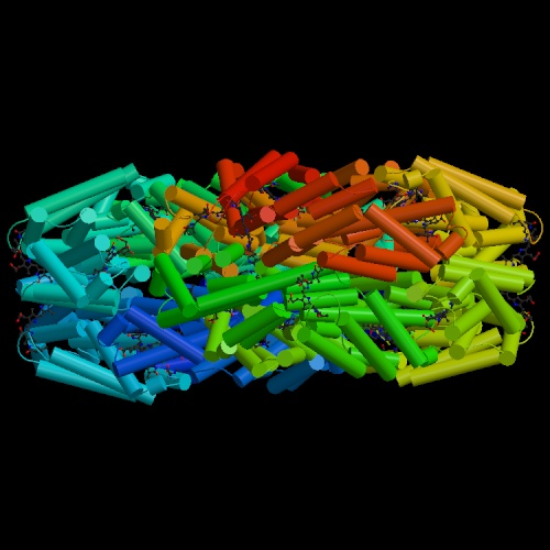
The tetrapyrrole units function as chromophores in this protein. To see them in the structure, make certain ligand is selected. I t may also be necessary to deselect water and change the appearance of the structure from ribbons to backbone, etc. In some viewers, the backbone can be deselected thus enabling you to see just the ligands and their arrangement relative to each other.
Below is a jpeg image of the protein. The rods represent the protein backbone. The linked five-membered rings in red and blue are the tetrapyrrole units.